By Krystal L. Beers, Certified Carnivore Nutrition Coach ©2017/2018
Canine Hip Dysplasia… it’s one of the Top 3 most talked about and controversial issues in canine health. It was first diagnosed in 1935. Since then generation upon generation of breeding dogs have been tested and selected for clear hips. Hip screening today is being aggressively pushed across the board of dog breed clubs, some going so far as to consider a breeder not “in good standing” if they don’t subject their dogs to radiograph tests.
Popular myth still dictates that it is purely a genetic factor, so only dogs with ‘Good’, ‘Excellent’, or the equivalent hip scores should be used for breeding. Yet here we are over 40 years later and Canine Hip Dysplasia (CHD) still has not been eliminated. Worse yet, two dogs certified ‘Excellent’ can still produce a pup with dysplastic hips. Have we been misled into performing unnecessary tests, tests that actually harm our dogs? There must be more to the story. As it turns out there is.
There are actually several factors that play a large role in the development of CHD, even though some will vehemently disagree. Those factors are: structure, nutrition, weight, surfaces, growth patterns, exercise, and hormones.
Chew on this…
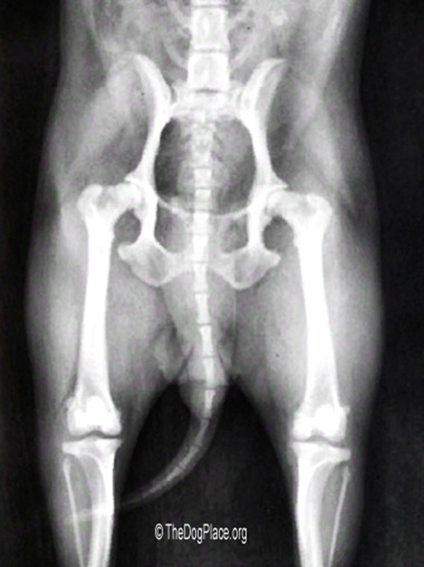
Dr. Wayne Riser, DVM founder of the Orthopedic Foundation for Animals (OFA) circa 1975, studied Canine Hip Dysplasia and explained it this way: “In all mammalian embryos, the hip is laid down as a single unit from mesenchymal tissue, and it develops normally as long as the components are left in full congruity. The hip is normal at some time in the development of the mammal, and abnormal development occurs only when stresses pull the components apart.
“In the dog, the hip is normal at birth. Intrauterine stresses are not sufficient to produce incongruity of the hip. The first time such forces are great enough is when the pup begins to take its position to nurse.
“Observations of the disease in man, dog, and a number of other mammals for many years have culminated in the conviction that the bony changes of hip dysplasia, regardless of species, occur because the soft tissues do not have sufficient strength to maintain congruity between the articular surfaces of the femoral head and the acetabulum.”
In a recent Natural Rearing Breeders Association (NRBA) newsletter, Dr. Jeannie Thomason, VND describes CHD in simpler terms as “a weakness in the ligaments that support the hip joint, allowing the ball of this ball-in-socket joint to bang against the joint surface, preventing the socket from forming properly. Instability created by weak supportive ligaments keeps the body from being able to manufacture a deep, smooth hip socket for the ball to fit snuggly into, resulting in the flattening of the acetabulum (hip socket) and a squaring of the femoral head (the ball).”
Which leads to the big question, can CHD be prevented? This article will explore each of the above mentioned factors involved in Canine Hip Dysplasia.
STRUCTURE
To begin with, instead of looking for terms such as “x-ray clear” of the OFA and PennHIP numbers, look to the structure of the breeding dogs. A key to preventing Canine Hip Dysplasia is found the descriptive words used that many dog breeders consider virtues: “big bone”, “huge feet”, “massive head”, etc. Those terms indicate excessive size and skeletal mass too dense for canines. We want moderation!
Look at wild dogs, wolves, coyotes, and foxes to see examples of canines properly supported by balanced frame and muscle mass that works without structural problems.
There are many books and videos on canine structure, even seminars. Become an expert in your own right, for your chosen breed. For those of you who are breeders, this will also help you in placing puppies.
NUTRITION
Before conception, nutrition the parents receive is vital, especially for the dam. Once mating occurs, if her body isn’t properly nourished with whole food protein and bioavailable vitamins/minerals, then the developing embryos will be adversely affected. The single most effective way to achieve excellent nutrition is through a balanced, whole food, Species Appropriate Raw Food diet.
Again, look to the wild relatives of the domestic dog as an example – raw meat, numerous organs, bones, marrow, even hooves and hides. The local wolf pack predating on free ranging cattle leaves behind only the lower jaw bone and tail switch. A lot of food for thought in that lesson!
PUPPY WEIGHTS
We’ve all seen those adorable photos of cute, roly-poly puppies! Few are aware that those puppies are most likely overweight. It is common knowledge that excess weight adversely affects joints, as well as other areas of health.
Yet this is a problem that often begins at weaning age when young puppies are started on kibble. Of course, we advocate a balanced Species Appropriate Raw Food diet (SARF), but what about when someone chooses to feed the commercial route. Most puppy foods contain abnormally high levels of protein, fat and calcium. Puppies need to grow more slowly than what such ingredients encourage.
Even brands that are fairly balanced are still a cooked product. Cooking/heat drastically changes the molecular structure of protein into what are called ‘polypeptides’. These partially broken down proteins become something the body does not recognize as food but as toxic foreign invaders the body needs to work very hard to remove. The immune system targets polypeptides, focusing energy on protecting itself against something that was eaten instead of on other areas that may need immune support, causing the entire system to work and perform extremely inefficiently. In turn, these undigested proteins lead to allergies and other diseases.
THE WHELPING BOX
The next contributing factor is the whelping area. Canine Hip Dyspasia starts in the whelping box, not in the parent’s hip scores. If you’ll recall in Part 1, we quoted Dr. Wayne Riser, DVM founder of the Orthopedic Foundation for Animals (OFA): “In the dog, the hip is normal at birth. Intrauterine stresses are not sufficient to produce incongruity of the hip. The first time such forces are great enough is when the pup begins to take its position to nurse.”
The best genetic structure and muscle strength can be ruined when very young puppies are on a slick surface like the commonly used kiddie pool. Even the wood box we use is slippery, so we add layers of various materials for traction. Some breeders on the web are posting about experiments with a ‘nest’ of sorts, trying to mimic the depression a mother creates in an earthen den so as to help young whelps move more naturally. The results are promising, even if the technique has not yet been perfected.
As soon as they step out of the nest to explore, litters of even toy breeds need to be allowed out in the natural environment for stable footing. And boy do they get around! It’s heartwarming to watch them grow stronger and faster every day engaging in this activity.
APPROPRIATE EXERCISE
Most people can hardly wait for their new family member to accompany them on excursions out-of-doors! Exercise is very important; however there can be too much of a good thing. When exercise is too much, inappropriate, and at too young of an age it carries negative consequences on the developing puppy’s joints – elbow and hip dysplasia, shoulder injuries – to name a few.
What is inappropriate exercise and how much? I’m glad you asked. This chart from Puppy Culture will give you a good idea:
HORMONE PRODUCTION
Hormones play a vital role in the proper growth of literally any species, humans included. Yet puppies are usually sexually altered by 6 months of age, and sometimes as young as 6 weeks old.
As we learned before, hormones do not signal the growth plates to close until 18 months of age. Abruptly cutting off hormone production long before then has a definite negative impact on normal and complete development of joints, ligaments, muscles, and tendons.
Most of you know we no longer perform elective joint X-rays on our dogs, nor do we use them during pregnancy to determine how many puppies a female will have. This is due to the health risks involved, namely infertility and cancer, and the gross lack of studies on their effects in animals. Most of the statistical information available is based on human studies.
WHAT ARE X-RAYS?
There are many different types of radiation – from the sun’s light to the heat coming off our bodies. But when talking about radiation and cancer risk, it is mostly man-made radiation from X-rays and gamma rays people think about.
X-rays and gamma rays can come from natural sources such as radon gas, radioactive elements in the earth, and cosmic rays that hit the earth from space. X-rays and gamma rays are types of high energy (high frequency) electromagnetic radiation. They are packets of energy that have no charge or mass (weight).
The American Cancer Society (ACS) explains that X-rays and gamma rays are both forms of ionizing radiation, which means they have enough energy to remove an electron from (ionize) an atom or molecule. Ionized molecules are unstable and quickly undergo chemical changes. If ionizing radiation passes through a cell in the body, it can lead to mutations (changes) in the cell’s DNA, the part of the cell that contains its genes (blueprints), causing germ-line mutations potentially affecting future generations. Caustic free radicals are also generated.
EXPOSURE RISKS
Sometimes the effect causes the cell to die, but sometimes it can lead to cancer later on. The amount of damage caused in the cell is related to the dose of radiation it receives. The damage takes place in only a fraction of a second, but other changes such as the beginning of cancer may take years to develop. The ACS flatly states: “Yes. X-rays and gamma rays are known human carcinogens (cancer-causing agents). The evidence for this comes from many different sources… Most studies on radiation and cancer risk have looked at people exposed to high doses of radiation… It is harder to measure the much smaller increase in cancer risk that might come from much lower levels of radiation exposure… Still, most scientists and regulatory agencies agree that even small doses of gamma and x-radiation increase cancer risk.”
FDA.gov acknowledges this information as well and goes on to note that the risk of developing cancer from medical imaging radiation exposure is generally very small, and is influenced by four things: 1) Radiation dose – the lifetime risk of cancer increases the larger the dose and the more X-ray exams a patient undergoes; 2) Patient’s age – the lifetime risk of cancer is larger for a patient who receives X-rays at a younger age than for one who receives them at an older age; 3) Patient’s sex – women are at a somewhat higher lifetime risk than men for developing radiation-associated cancer after receiving the same exposures at the same ages; 4) Body region – some organs are more radiosensitive than others.
According to radiologyinfo.org, one radiograph of the lower G.I. tract in an adult human exposes them to the same amount of radiation that would be naturally absorbed (from the environment) over a 3 year period. To have a dogs hips certified requires three x-ray views to be taken. In 2014 studies showed that the lifetime risk of fatal cancer from that single G.I. x-ray exposure is “low” compared to a CT scan which is “moderate.” (Risk vs. Benefit philosophy- so how much according to whom?! Each individual organism has different thresholds for each and every exposure.)
For children, exposure to radiation from imaging tests is of particular concern, because children are still growing and much more sensitive to radiation than adults. The ACS says this means that for a young child, the risk of developing a radiation-related cancer could be several times higher than for an adult exposed to the same imaging test. The risks from these tests are not known for sure, but to be safe, most doctors recommend that children only get these tests when they are absolutely needed. When such tests are done, it is important to use the minimum amount of radiation needed to get the image.
The FDA has urged makers of medical scanning equipment to make new devices that would minimize the radiation dose delivered to children, and goes on to say “it’s important to use the lowest dose possible with scanning a child. But it’s even more important to avoid unnecessary scans in the first place.”
What about our dogs? Are there precautions in place for them, too?
Ponder this from TheDogPlace.org: A mastiff having an abdominal or hip X-ray would get a 6 year dose of radiation – a Corgi about a 25 year dose. If one year of our life is equivalent to about seven years in a dog’s, does that mean a radiograph X-ray is seven times more dangerous for your dog than for you?
Kevin S. Toppenberg, M.D., D. Ashley Hill, M.D., and David P. Miller, M.S. of the Florida Hospital Medical Center, Orlando, Florida wrote that the most sensitive time period for central nervous system teratogenesis for a human fetus is between 10 and 17 weeks of gestation. Non-urgent radiologic testing should be avoided during this time. Rare consequences of prenatal radiation exposure include a slight increase in the incidence of childhood leukemia and, possibly, a very small change in the frequency of genetic mutations.
Is the “most sensitive” time period for canine fetuses known?
Several national and international agencies study different substances in the environment to determine if they can cause cancer. Based on lab animal and human evidence, several expert agencies have evaluated the cancer-causing nature of X-rays and gamma rays.
The International Agency for Research on Cancer (IARC) is part of the World Health Organization (WHO). Its major goal is to identify causes of cancer. Based on the data available, IARC classifies x- and gamma radiation as a “known human carcinogen.” The National Toxicology Program (NTP) is formed from parts of several different US government agencies, including the National Institutes of Health (NIH), the Centers for Disease Control and Prevention (CDC), and the Food and Drug Administration (FDA). The NTP has classified x- and gamma radiation as “known to be a human carcinogen.” The US Environmental Protection Agency (EPA) sets limits for exposure to x-rays and gamma rays because it recognizes that this form of radiation can cause cancer.
Is the stark rise of cancer in dogs due in part to the cumulative and generational damage of X-rays?
ORGAN SHIELDING
But don’t they shield my dog’s reproductive organs? The X-ray has to be unobstructed and strong enough to reveal bone and joint density. It doesn’t take a brain surgeon to understand this means the ovaries and testicles receive a whopping dose of radiation.
Case in point: My husband needed X-rays for spinal record for chiropractor treatment. The radiologist had to strengthen power for a through chest view, thus double exposure for the one view. It was explained that the mass of the body demanded higher exposure for the view to be readable. So, dosage is variable in each instance controlled by the technician and equipment.
It is now well known that pharmaceutical and pet food companies provide “teaching materials” to veterinary universities, no doubt containing their own slant and agenda. It only stands to reason then that most freshly minted animal doctors will continue to disregard the cumulative effects of radiation. And beaming powerful radiation directly at our dog’s reproductive organs won’t trouble them just as it hasn’t troubled their predecessors.
What are the cumulative exposure risks for a dog over its lifetime? How much radiation is too much? Unfortunately we will likely never know the answer to those pertinent questions. In this case it would be prudent to err on the side of caution.
I fervently hope this article has given you a different perspective on Canine Hip Dysplasia, the many factors involved in its development and prevention. As well as proffered an enlightened view of elective X-rays so that you can make informed decisions about radiation risks for your dogs, and your own health.


Leave A Comment